 Editorial NexoScience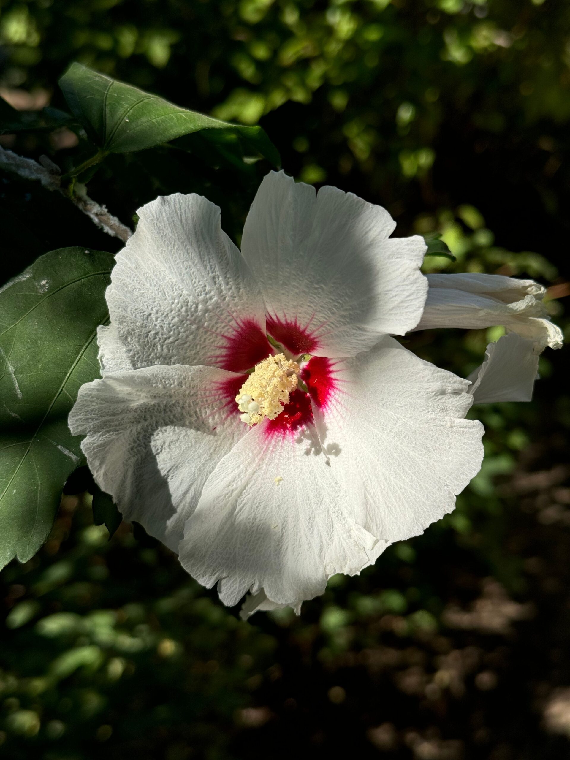
Look into the center of a Hibiscus trionum flower on a bright morning and the dark purple base of each petal throws back a faint metallic sheen, the same oily blue you notice on a soap bubble or the shell of a beetle. That sheen is not a pigment at all, and a team at the University of Cambridge has now shown that the plant decides where to place it by controlling the chemical recipe of its own surface layer, not simply by growing faster or thicker.
The blue halo comes from microscopic ridges, not from color molecules. The base of the petal is covered with parallel striations in the cuticle, the waxy polymer coating every aerial surface of a plant. Those ridges are only semi ordered, and that imperfection matters: the disorder scatters light toward the blue and ultraviolet end of the spectrum, producing a halo that bumblebees can detect and that improves their foraging efficiency in laboratory tests. Physicists had already explained how the optical effect works. How a growing cell manufactures a grating that regular was, in the authors own words, obscure.
The simplest explanation did not survive the experiments. An earlier model proposed that the pattern followed from just two numbers, how much the cell stretches and how fast cuticle is produced, with the mismatch between them buckling the surface into ridges. The team dosed developing buds with hormones and cytoskeleton drugs, including IAA, gibberellic acid, NPA, oryzalin and Taxol, which changed cell dimensions but never removed the striations. They then compared eight species and accessions of Hibiscus, five striated and three smooth. In the smooth ones, the measured growth and cuticle values predicted ridges that simply were not there.
Interfering with the chemistry switched the pattern off. Working in a species with a seven week cycle from seed to flower, the researchers built a transformation protocol and made transgenic lines. One gene, HtSHINE3, is expressed only in the pigmented region and rises roughly two hundredfold above its relatives as the ridges appear. Altered versions of it produced flowers with no blue sheen and a completely smooth petal base, even though Fat Red staining and CryoSEM fractures showed the same cell shape, cuticle thickness and cuticle architecture as wild type. Disrupting cutin assembly or wax production, using AtCDEF1, HtCUS1, HtMIXTA-like1 and AtDEWAX, degraded the grating in different ways, leaving faint, interrupted or misaligned ridges.
Striated surfaces share a chemical fingerprint. Using LESA-MS, a technique that samples only the outermost surface, the team profiled cuticles from transgenic lines and from the different species. A principal component analysis separated striated from smooth samples along a single axis. Striated cuticles were associated with free dihydroxy palmitic acid, a key cutin monomer, and with very long chain waxes, while smooth ones tended to carry probable phenolic compounds. Mechanical tests closed the loop: pressing on immature wild type petals forced ridges to appear, while the same force applied to chemically altered petals produced almost nothing.
This is a mechanism in one model system, not a law of botany. The chemical profiles come from associations in a statistical analysis, and the direct causal evidence rests on transgenic manipulation in a single laboratory species. What the work establishes is that a plant can tune cuticle chemistry with cell by cell precision so that the same physical stress buckles one region and leaves the neighboring one flat. For anyone trying to imitate structural color in synthetic materials, that is the interesting part, because it suggests the pattern is written into the material rather than imposed from outside.
Source: “Cuticle chemistry drives the development of diffraction gratings on the surface of Hibiscus trionum petals”, by Edwige Moyroud, Chiara A. Airoldi and colleagues, with Beverley J. Glover as senior author, published in Current Biology (2022), volume 32, pages 5323 to 5334. DOI: 10.1016/j.cub.2022.10.065. Open access under a CC BY 4.0 license.