 Editorial NexoScience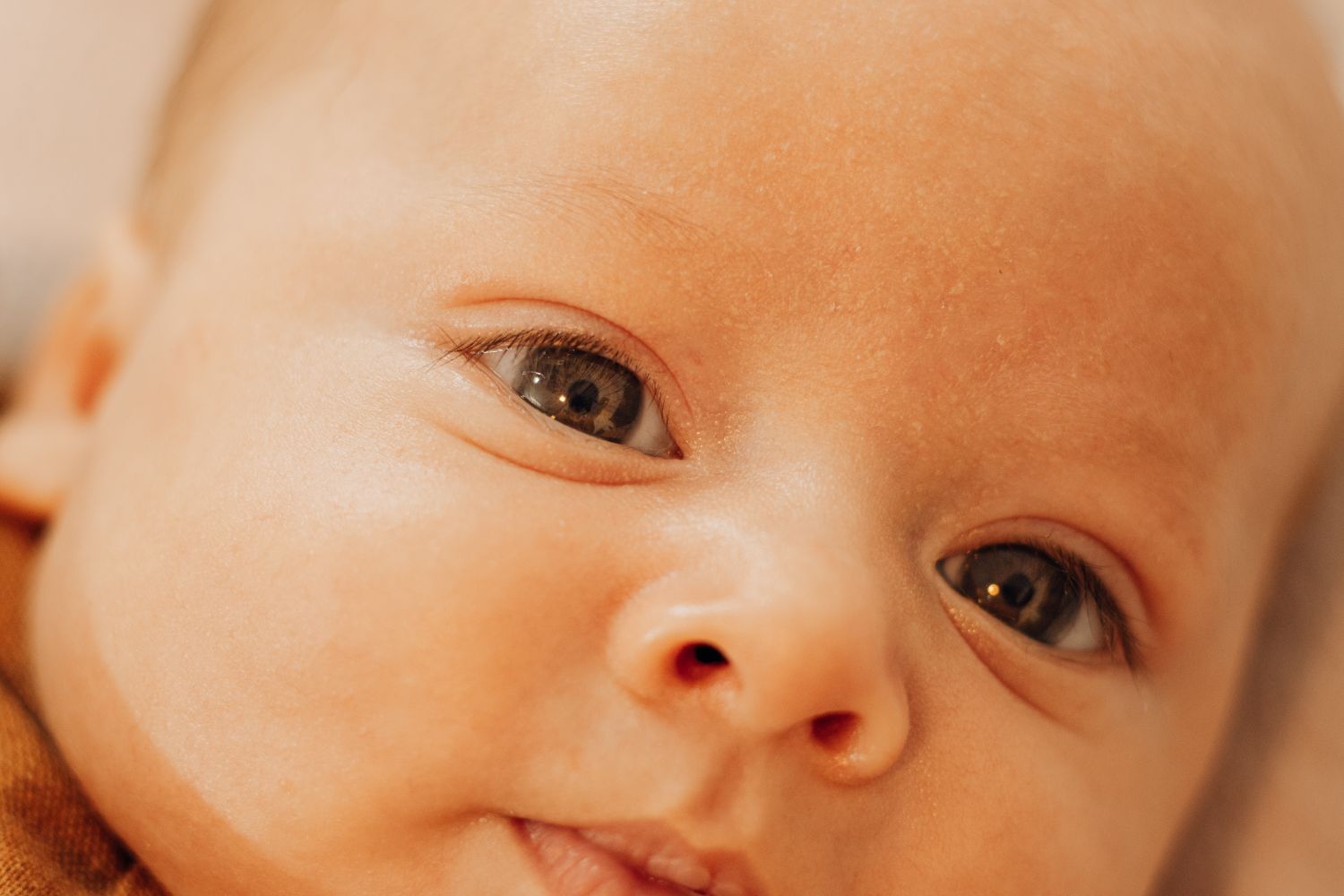
Doctors once read a newborn’s face like a wiring diagram. For years, the branches of the trigeminal nerve, the large facial nerve whose lower branch a dentist numbs before drilling, gave pediatricians what looked like a ready-made grid: one branch across the forehead, one across the cheek, one across the jaw. Where a red birthmark landed on that grid was how a baby’s risk of a rare brain disorder got judged, and whether that baby was sent for a scan. The grid turned out to be the wrong tool for the job.
The correction is now written into pediatric guidance in plain language. A review published in September 2022 in the journal Pediatrics in Review states that the V1, V2 and V3 nerve zones are no longer used to describe where a port-wine birthmark sits, because the marks have proved not to be related to the trigeminal nerve in any capacity. They are vascular in origin, and their outline on the skin follows how blood vessels developed in the embryo. The authors, dermatologists and an ophthalmologist at Baylor College of Medicine, Nationwide Children’s Hospital, the University of California, Irvine, and Texas Children’s Hospital, wrote it as an update for the pediatricians who see these babies first.
The disorder they are screening for is Sturge-Weber syndrome. It is a neurocutaneous condition, meaning it hits skin and nervous system together, and it classically appears as a triad of vascular malformations in the skin, the eyes and the brain. A diagnosis usually requires at least two of three findings: a characteristic facial port-wine birthmark, malformed vessels in the eye, and MRI evidence of leptomeningeal angiomatosis, a tangle of abnormal vessels draped over the surface of the brain. Children with brain involvement can develop epilepsy, strokelike episodes, headaches, developmental delays and encephalopathy, and anxiety and depression are common alongside them.
The birthmark and the brain share a plumbing system, not a nerve. Both the birthmark and the syndrome usually trace back to a somatic activating mutation in GNAQ, a genetic change that arises in the affected tissue itself rather than being inherited from a parent. It drives a growth pathway that blocks cells from dying off on schedule, and vessels keep building where they should have stopped. Crucially, the skin of the forehead grows out of the same vascular territory as the cerebral cortex and the eye, which is why those three organs turn up affected as a set. Judging the risk by nerve branches was a bit like predicting where a river will flood by consulting the subway map that runs above it. Two networks over the same ground, and only one of them carries the water.
The forehead is the single best independent predictor. For this purpose the forehead is defined generously, with its lower border drawn from the outer corner of the eye to the top of the ear, eyelid included. Within that zone, two patterns raise the alarm: a mark off to one side that covers more than half of that side of the forehead, or a mark anywhere on the midline, however small. A comparable mark covering less than half of one side of the forehead is considered low risk for the syndrome. Risk also climbs with reach, so a birthmark that spreads across an entire half of the face, or across both sides, carries a higher risk than one that stops at the forehead.
A birthmark on the eyelid carries a separate risk that never expires. Any mark involving the skin around the eye, upper or lower lid, puts that child at risk of acquired glaucoma, damaging pressure inside the eye, and that means scheduled eye examinations for life even when the first screens come back clean. An eye examination in the newborn period is urgent, because the glaucoma linked to this syndrome can be present at birth. The authors note the reverse case as well: an infant with an upper eyelid birthmark that does not touch the forehead is at low risk for brain involvement, and still needs the eye monitoring.
Most red marks on a newborn’s forehead are something else entirely. By far the commonest look-alike is nevus simplex, the mark parents know as an angel’s kiss, and telling the two apart is most of the job. Nevus simplex is lighter, patchier and blurry at the edges, usually comes with matching marks on the eyelids, the philtrum or the nape, and typically fades over the first year. A port-wine birthmark is a solid, sharply outlined patch that never involutes and grows in proportion with the child. Two other conditions can also be mistaken for it early on: a segmental infantile hemangioma, which starts to proliferate and become raised within a few weeks of birth, and an arteriovenous malformation, which over time turns warm, swollen and pulsatile. That is why the review sends uncertain cases to a vascular birthmark specialist before anyone orders a workup for the syndrome.
The laser works best before a baby’s first birthday. Pulsed dye laser is the gold standard treatment, targeting the hemoglobin inside the birthmark’s vessels, and it has the longest safety record in children of any laser used for this. Starting in infancy stacks the odds: the skin is thinner, the birthmark is thinner, its vessels are smaller and closer to the surface, and there is less melanin competing for the laser’s light. Treatment is a commitment rather than an appointment, with a series that runs anywhere from fewer than five sessions to thirty or more, spaced a month or so apart. Early treatment also tends to spare the child general anesthesia, and gets the initial series finished before school starts, which is as much a psychosocial calculation as a dermatological one.
The laser lightens the birthmark, it does not erase it. Parents are told this up front, because even after a successful series the redness usually rebounds and maintenance sessions are expected for life. The procedure leaves purpura, bruise-like discoloration that is usually painless but more conspicuous than the birthmark itself, for a couple of weeks afterward, and it carries small risks of blistering, temporary pigment change and, rarely, scarring. Those last two risks are higher in children with more pigmented skin, since melanin absorbs the laser light too. Left alone, a facial birthmark tends to deepen from red toward purple, thicken, and develop fragile bumps that bleed, usually from puberty onward, and treating early can head off at least some of that progression.
The review is a synthesis of the field, not an experiment, and it labels itself as one. Its own summary marks each recommendation by the strength behind it, separating the points that rest on recent research evidence, such as the forehead rule, from the ones that rest on expert consensus, such as the referral pathway. That distinction matters, because the forehead rule describes a pattern observed in children who have the syndrome rather than a mechanism anyone tested here, and the shared embryonic origin of forehead skin, eye and cortex is the proposed explanation for that pattern, not proof of it. A high-risk birthmark is a reason to look, not a diagnosis, and the syndrome has been reported, rarely, in children with no birthmark at all. The authors are also openly candid about the one decision they cannot settle: when to do the brain MRI. There is no consensus, they wrote, because a normal scan early in infancy can be falsely negative and so cannot rule anything out, while scanning early is what would make preventive treatment with antiseizure medication or aspirin possible in the first place. Anesthesia risk sits on the other side of that ledger, although some experienced centers now image young infants awake, fed and swaddled into sleep.
The first thing the authors want parents told is that they did not cause this. Guilt is close to universal in these families, over the birthmark and over the decision to pursue laser treatment or skip it, and the review is blunt that nothing a parent did or failed to do produced either the mark or the syndrome. What comes next is aimed one level deeper than the skin. Pulsed dye laser reaches only about a twentieth of an inch (1.2 millimeters) into the skin, missing the deeper vessels, and the lasers that go deeper scar. So the authors put their optimism in imaging that could map the size and depth of each birthmark’s vessels from the surface before a single pulse is fired, and in drugs aimed at GNAQ itself, following the gene-targeted therapies already in use in oncology and now reaching other vascular birthmarks.
Source: Poliner A, Fernandez Faith E, Blieden L, Kelly KM, Metry D. “Port-wine Birthmarks: Update on Diagnosis, Risk Assessment for Sturge-Weber Syndrome, and Management.” Pediatrics in Review, 2022;43(9):507-516. DOI: 10.1542/pir.2021-005437. Open access.
This article summarizes published scientific research for general information. It is not medical advice and does not replace consultation with a qualified health professional.